Panorex
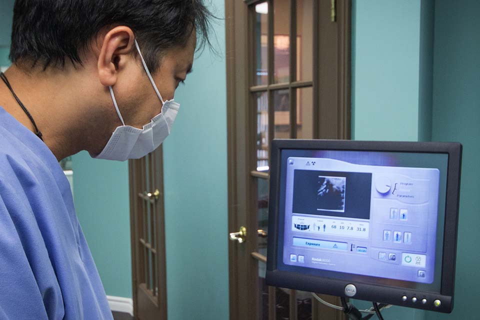
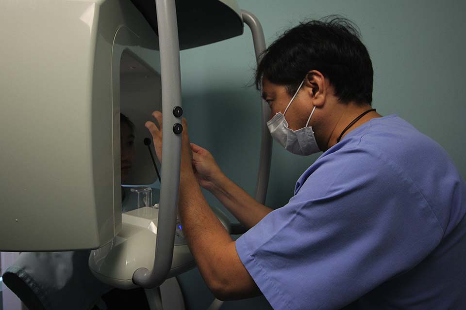
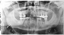
A panoramic radiograph is a panoramic scanning dental X-ray of the upper and lower jaw. It shows a two-dimensional view of a half-circle from ear to ear. Panoramic radiography is a form of tomography; thus, images of multiple planes are taken to make up the composite panoramic image, where the maxilla and mandible are in the focal trough and the structures that are superficial and deep to the trough are blurred.
Panorex can be used to provide information on:
- Impacted wisdom teeth diagnosis and treatment planning – the most common use is to determine the status of wisdom teeth and trauma to the jaws.
- Periodontal bone loss and periapical involvement.
- Finding the source of dental pain
- Assessment for the placement of dental implants
- Orthodontic assessment. pre and post operative
- Diagnosis of developmental anomalies such as cherubism, cleido cranial dysplasia
- Carcinoma in relation to the jaws
- Temporomandibular joint dysfunctions and ankylosis.
- Diagnosis of osteosarcoma, ameloblastoma, renal osteodystrophy affecting jaws and hypophosphatemia.
- Diagnosis, and pre- and post-surgical assessment of oral and maxillofacial trauma, e.g. dentoalveolar fractures and mandibular fractures.
- Salivary stones (Sialolithiasis).
- Other diagnostic and treatment applications.

IntraOral Camera
IntraOral Cameras (“IOCs”) are used to show you a clear picture of the inside of your mouth, allowing the dentist to consult on various treatment options, and save the images directly to your file.
As a patient, you can see your own intraoral conditions under magnification with better quality than the dentist could show you with a foggy mouth mirror.


